Click image to see more details
Product Info Summary
| SKU: | PA1503 |
|---|---|
| Size: | 100 μg/vial |
| Reactive Species: | Human |
| Host: | Rabbit |
| Application: | WB |
Customers Who Bought This Also Bought


Product info
Product Name
Anti-MCL1 Antibody
SKU/Catalog Number
PA1503
Size
100 μg/vial
Form
Lyophilized
Description
Boster Bio Anti-MCL1 Antibody catalog # PA1503. Tested in WB applications. This antibody reacts with Human.
Storage & Handling
Store at -20˚C for one year from date of receipt. After reconstitution, at 4˚C for one month. It can also be aliquotted and stored frozen at -20˚C for six months. Avoid repeated freeze-thaw cycles.
Cite This Product
Anti-MCL1 Antibody (Boster Biological Technology, Pleasanton CA, USA, Catalog # PA1503)
Host
Rabbit
Contents
Each vial contains 5mg BSA, 0.9mg NaCl, 0.2mg Na2HPO4, 0.05mg Thimerosal, 0.05mg NaN3.
Clonality
Polyclonal
Isotype
Rabbit IgG
Immunogen
A synthetic peptide corresponding to a sequence at the C-terminus of human MCL1, different from the related mouse sequence by one amino acid.
*Blocking peptide can be purchased. Costs vary based on immunogen length. Contact us for pricing.
Cross-reactivity
No cross-reactivity with other proteins
Reactive Species
PA1503 is reactive to MCL1 in Human
Applications
PA1503 is guaranteed for WB Boster Guarantee
Observed Molecular Weight
37 kDa
Calculated molecular weight
37.337kDa
Background of Mcl-1
BCL2L3, also known as MCL1 (myeloid cell leukemia sequence 1) encodes an anti-apoptotic protein, which is a member of the Bcl-2 family. Alternative splicing results in multiple transcript variants. The longest gene product (isoform 1) enhances cell survival by inhibiting apoptosis while the alternatively spliced shorter gene products (isoform 2 and isoform 3) promote apoptosis and are death-inducing. Using the methods of somatic cell hybrid analysis and fluorescence in situ hybridization), the MCL1 gene is mapped to human 1q21.MCL1is a critical and specific regulator essential for ensuring the homeostasis of early hematopoietic progenitors. Phosphorylation of MCL1 directs its interaction with the tumor suppressor protein FBW7, which is the substrate-binding component of a ubiquitin ligase complex. The polyubiquitylation of MCL1 then targets it for proteasomal degradation. The degradation of MCL1 was blocked in patient-derived tumor cells that lacked FBW7 or had loss-of-function mutations in FBW7, conferring resistance to antitubulin agents and promoting chemotherapeutic-induced polyploidy.
Antibody Validation
Boster validates all antibodies on WB, IHC, ICC, Immunofluorescence, and ELISA with known positive control and negative samples to ensure specificity and high affinity, including thorough antibody incubations.
Innovating Scientists Reward
If you are the first to review this product, or if you have results for a special sample, species or application this product is not validated in, share your results with us and receive product credits you can use towards any Boster products! Applicable to all scientists worldwide.
Submit A Review
Assay dilution & Images
Reconsitution
Add 0.2ml of distilled water will yield a concentration of 500ug/ml.
Assay Dilutions Recommendation
The recommendations below provide a starting point for assay optimization. The actual working concentration varies and should be decided by the user.
Western blot, 0.1-0.5μg/ml, Human
Validation Images & Assay Conditions
Click image to see more details
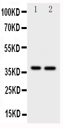
Anti-MCL1 antibody, PA1503, Western blotting
All lanes: Anti MCL1 (PA1503) at 0.5ug/ml
Lane 1: HELA Whole Cell Lysate at 40ug
Lane 2: MCF-7 Whole Cell Lysate at 40ug
Predicted bind size: 37KD
Observed bind size: 37KD
Protein Target Info & Infographic
Gene/Protein Information For MCL1 (Source: Uniprot.org, NCBI)
Gene Name
MCL1
Full Name
Induced myeloid leukemia cell differentiation protein Mcl-1
Weight
37.337kDa
Superfamily
Bcl-2 family
Alternative Names
BCL2L3; bcl2-L-3; BCL2L3MGC104264; Bcl-2-like protein 3; Bcl-2-related protein EAT/mcl1; EAT; induced myeloid leukemia cell differentiation protein Mcl-1; Mcl1; Mcl-1; mcl1/EAT; MCL1-ES; MCL1L; MCL1S; MGC1839; myeloid cell leukemia ES; myeloid cell leukemia sequence 1 (BCL2-related); TM MCL1 BCL2L3, EAT-ES, MCL1L, MCL1S, Mcl-1, TM, bcl2-L-3, mcl1/EAT, MCL1 MCL1 apoptosis regulator, BCL2 family member induced myeloid leukemia cell differentiation protein Mcl-1|BCL2 family apoptosis regulator|MCL1, BCL2 family apoptosis regulator|bcl-2-like protein 3|bcl-2-related protein EAT/mcl1|myeloid cell leukemia 1|myeloid cell leukemia ES|myeloid cell leukemia sequence 1 (BCL2-related)
*If product is indicated to react with multiple species, protein info is based on the gene entry specified above in "Species".For more info on MCL1, check out the MCL1 Infographic

We have 30,000+ of these available, one for each gene! Check them out.
In this infographic, you will see the following information for MCL1: database IDs, superfamily, protein function, synonyms, molecular weight, chromosomal locations, tissues of expression, subcellular locations, post-translational modifications, and related diseases, research areas & pathways. If you want to see more information included, or would like to contribute to it and be acknowledged, please contact [email protected].
Specific Publications For Anti-MCL1 Antibody (PA1503)
Hello CJ!
PA1503 has been cited in 1 publications:
*The publications in this section are manually curated by our staff scientists. They may differ from Bioz's machine gathered results. Both are accurate. If you find a publication citing this product but is missing from this list, please let us know we will issue you a thank-you coupon.
Liu N, Chen T, Wang X, Yang D, Xue B, Zhu H. Febs Lett. 2015 Apr 2;589(8):897-903. Doi: 10.1016/J.Febslet.2015.02.026. Epub 2015 Mar 3. Msi1 Confers Resistance To Trail By Activating Erk In Liver Cancer Cells.
Recommended Resources
Here are featured tools and databases that you might find useful.
- Boster's Pathways Library
- Protein Databases
- Bioscience Research Protocol Resources
- Data Processing & Analysis Software
- Photo Editing Software
- Scientific Literature Resources
- Research Paper Management Tools
- Molecular Biology Software
- Primer Design Tools
- Bioinformatics Tools
- Phylogenetic Tree Analysis
Customer Reviews
Have you used Anti-MCL1 Antibody?
Submit a review and receive an Amazon gift card.
- $30 for a review with an image
Be the first to review Anti-MCL1 Antibody
*The first user to submit a review for a product is eligible for Boster's Innovating Scientists Reward, which gives product credits. This is in addition to the gift card reward.
Customer Q&As
Have a question?
Find answers in Q&As, reviews.
Can't find your answer?
Submit your question
4 Customer Q&As for Anti-MCL1 Antibody
Question
We have observed staining in human thalamus. What should we do? Is anti-MCL1 antibody supposed to stain thalamus positively?
Verified Customer
Verified customer
Asked: 2020-05-01
Answer
According to literature thalamus does express MCL1. According to Uniprot.org, MCL1 is expressed in epithelium of mammary gland, myeloid leukemia cell, myeloid leukemia cell neuroblastoma, thalamus, mammary gland placenta, ewing sarcoma, cervix carcinoma, among other tissues. Regarding which tissues have MCL1 expression, here are a few articles citing expression in various tissues:
Cervix carcinoma, Pubmed ID: 18669648
Ewing sarcoma, Pubmed ID: 10634649
Mammary gland, and Placenta, Pubmed ID: 15489334
Myeloid leukemia cell, Pubmed ID: 7682708
Myeloid leukemia cell, and Neuroblastoma, Pubmed ID: 10766760
Thalamus, Pubmed ID: 14702039
Boster Scientific Support
Answered: 2020-05-01
Question
We were satisfied with the WB result of your anti-MCL1 antibody. However we have seen positive staining in myeloid leukemia cell membrane using this antibody. Is that expected? Could you tell me where is MCL1 supposed to be expressed?
Verified Customer
Verified customer
Asked: 2020-04-23
Answer
Based on literature, myeloid leukemia cell does express MCL1. Generally MCL1 expresses in membrane. Regarding which tissues have MCL1 expression, here are a few articles citing expression in various tissues:
Cervix carcinoma, Pubmed ID: 18669648
Ewing sarcoma, Pubmed ID: 10634649
Mammary gland, and Placenta, Pubmed ID: 15489334
Myeloid leukemia cell, Pubmed ID: 7682708
Myeloid leukemia cell, and Neuroblastoma, Pubmed ID: 10766760
Thalamus, Pubmed ID: 14702039
Boster Scientific Support
Answered: 2020-04-23
Question
I am interested in using your anti-MCL1 antibody for cellular homeostasis studies. Has this antibody been tested with western blotting on hela whole cell lysate? We would like to see some validation images before ordering.
Verified Customer
Verified customer
Asked: 2020-02-14
Answer
We appreciate your inquiry. This PA1503 anti-MCL1 antibody is tested on hela whole cell lysate. It is guaranteed to work for WB in human. Our Boster guarantee will cover your intended experiment even if the sample type has not been be directly tested.
Boster Scientific Support
Answered: 2020-02-14
Question
We are currently using anti-MCL1 antibody PA1503 for human tissue, and we are content with the WB results. The species of reactivity given in the datasheet says human. Is it likely that the antibody can work on bovine tissues as well?
Verified Customer
Verified customer
Asked: 2019-07-11
Answer
The anti-MCL1 antibody (PA1503) has not been tested for cross reactivity specifically with bovine tissues, but there is a good chance of cross reactivity. We have an innovator award program that if you test this antibody and show it works in bovine you can get your next antibody for free. Please contact me if I can help you with anything.
Boster Scientific Support
Answered: 2019-07-11
